Skip to content
Artwork for Quantum Imaging Lab

Quantum Imaging Lab

Sanjay Arya, M.S.R.T. (R)(MR), MRSO

Quantum Imaging Lab, presented by Professor Sanjay Arya, M.S., R.T.(R)(MR), provides structured, exam-aligned audio learning for radiologic technology students preparing for the ARRT exam and educators supporting instruction.

Episodes cover core ARRT Content Specification areas including Image Production, Radiation Protection, Safety, and Procedures through focused microlearning.

Content reflects topics commonly taught in radiologic technology programs and supports ARRT certification and Continuing Qualifications Requirements (CQR).

© 2026 Quantum Imaging Lab.

Play
  • 45 episodes
  • Avg 47 min
  • English
Counted on this page — what you have heard stays on this device, so it is not something the list can be paged by.
  • S1 · E5
    April 27 · 31 min

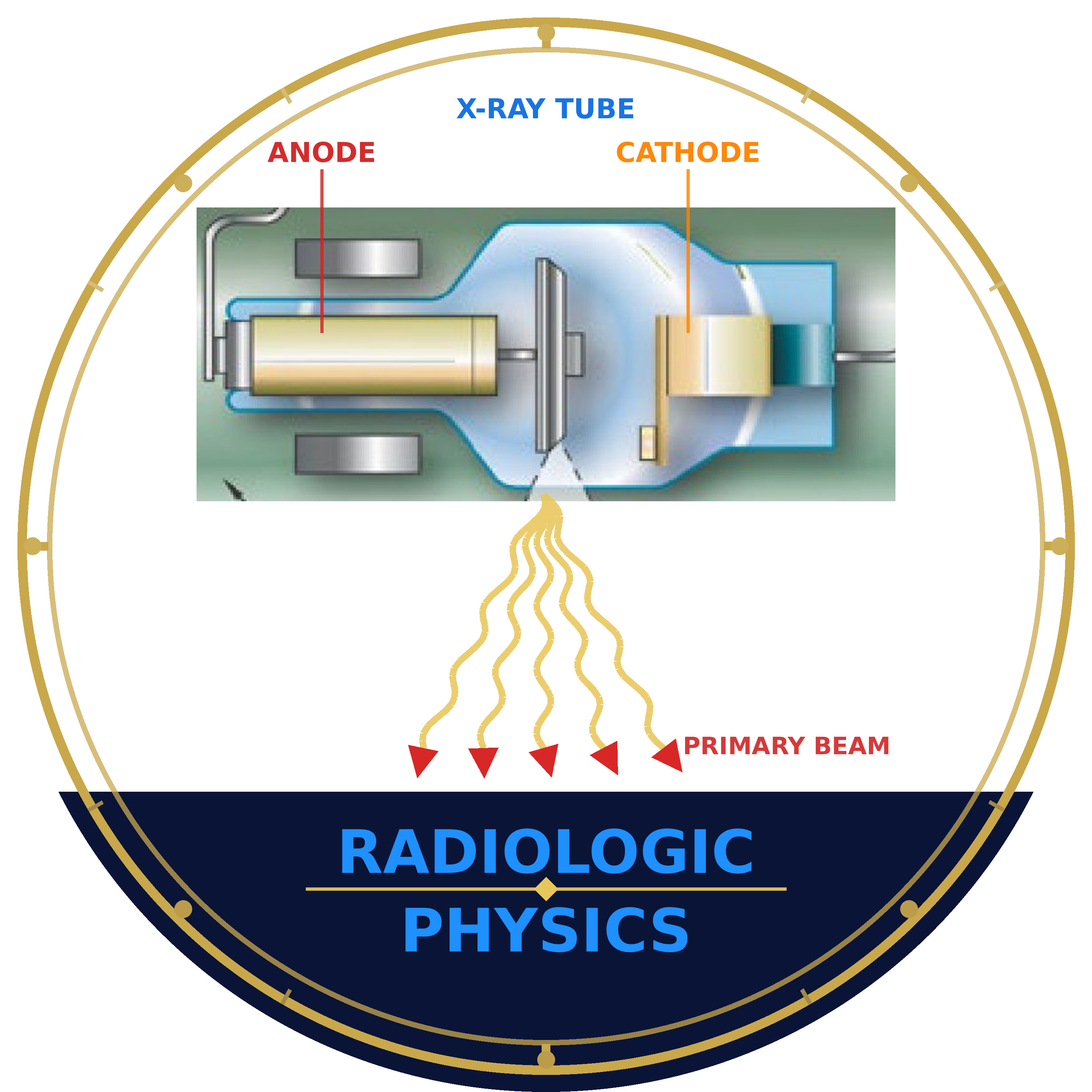
    [Radiologic Physics] Ep 5 – Inverse Square Law

    This episode opens with a review of wave-particle duality, establishing how x-rays can be described both as waves — characterized by wavelength and frequency — and as photons that interact with matter. Listeners explore how photon wavelength determines what size of matter it interacts with, from radio waves interacting with metal antennae down to x-ray photons interacting with atoms and electrons. Key terminology is introduced: radiopaque, radiolucent, attenuation, and intensity — along with the distinctions between reflection, transmission, attenuation, and absorption as x-rays and visible light pass through matter. The second half of the episode focuses on beam divergence and the Inverse Square Law. The cone-shaped geometry of the x-ray field is explained, showing why intensity decreases as photons spread over a larger area with increasing distance. The ISL formula (I₁/I₂ = D₂²/D₁²) is introduced and applied through worked clinical examples — including how doubling the SID reduces intensity to one-quarter and halving the distance quadruples it — with direct connections to SID adjustments, patient dose, and radiation protection practice. This episode aligns with the Safety content category — Radiation Physics and Radiobiology subcategory — of the ARRT Radiography Examination Content Specifications. Audio content is adapted from original instructional material developed by Professor Sanjay Arya, M.S., R.T.(R)(MR) for radiologic technology education. Part of the Radiologic Physics series — Quantum Imaging Lab. © 2026 Quantum Imaging Lab. All rights reserved.
  • S1 · E3
    April 27 · 41 min

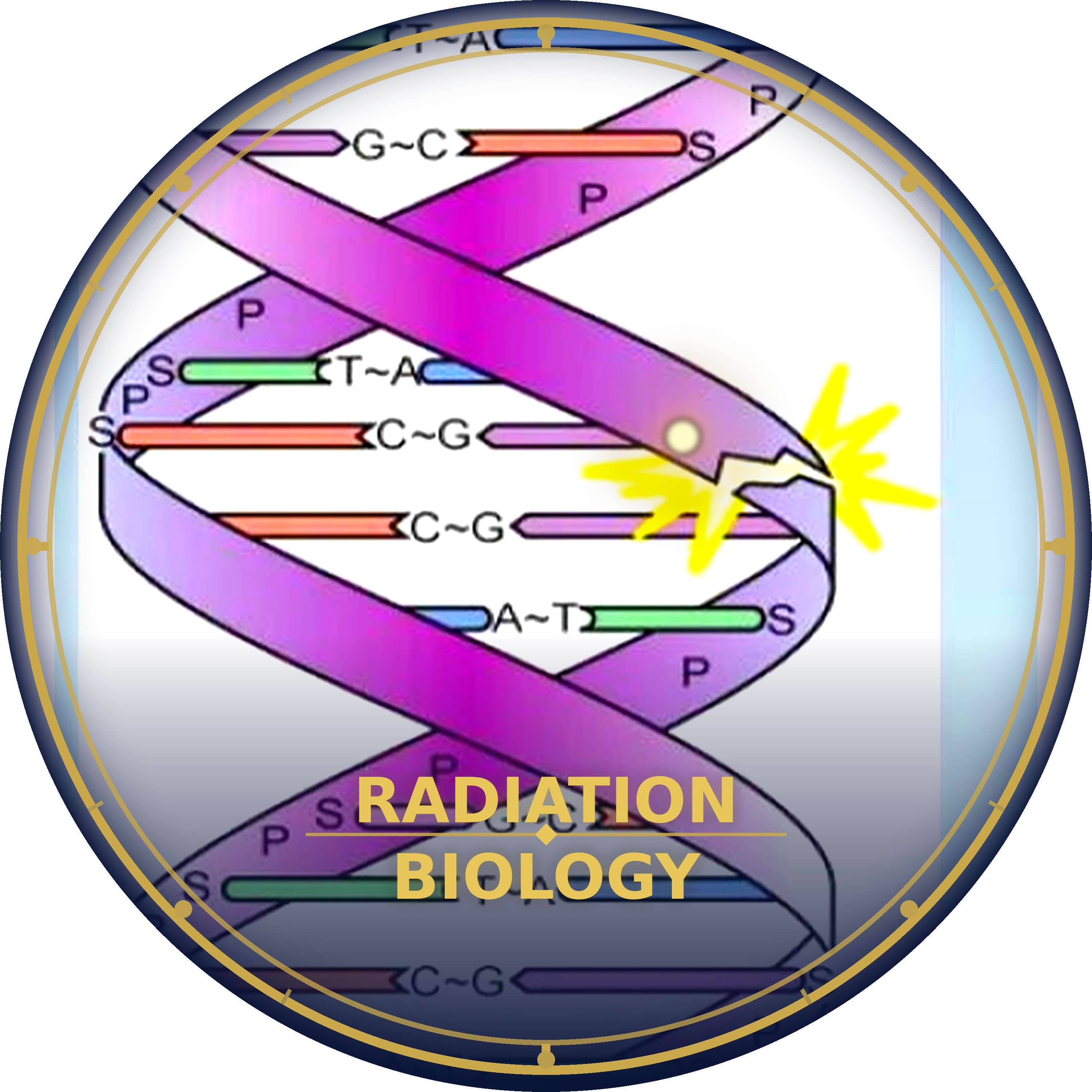
    [Radiation Biology] Ep 3 – Cell Structure, Function, and Division

    Cells are the fundamental building blocks of all living matter, and understanding their architecture is essential to grasping how radiation causes biological harm. This episode covers the structural organization of the human body from organ systems down to the individual cell, introduces cell theory, and explores the major components of the cell — including the plasma membrane, nucleus, and cytoplasmic organelles such as mitochondria, ribosomes, the endoplasmic reticulum, Golgi apparatus, and lysosomes. Special attention is given to the nucleus as the most radiation-sensitive component of the cell, and to the chemical composition of protoplasm including both inorganic and organic compounds. The episode takes a focused look at the four classes of organic molecules — carbohydrates, lipids, proteins, and nucleic acids — with particular emphasis on DNA and RNA. The double-helical structure of DNA, its base-pairing rules, and its role as the master chemical governing all cellular functions are examined alongside the three types of RNA and their roles in protein synthesis. The two-step process of transcription and translation is walked through in detail, connecting molecular biology directly to the radiation biology principle that DNA is the primary radiosensitive target molecule within the living cell. Content is structured to support radiologic technology programs preparing for imaging coursework and ARRT certification review. This episode aligns with the Safety content category — Radiation Physics and Radiobiology subcategory — of the ARRT Radiography Examination Content Specifications. Audio content is adapted from original instructional material developed by Professor Sanjay Arya, M.S., R.T.(R)(MR) for radiologic technology education. Part of the Radiation Biology series — Quantum Imaging Lab. © 2026 Quantum Imaging Lab. All rights reserved.

  • S1 · E4
    April 20 · 15 min

    [Radiologic Physics] Ep 4 – Electromagnetic Energy

    This episode provides a focused examination of electromagnetic energy and its properties as they apply to radiologic science. Radiation is defined broadly before narrowing to electromagnetic energy — its photon-based composition, massless and chargeless nature, sinusoidal travel at the speed of light, and three fundamental wave properties: wavelength, frequency, and amplitude. The inverse relationship between wavelength and frequency is established through the wave equation (c = λ × f), with a worked numerical example, and Planck's equation (E = hν) is introduced to connect photon energy directly to frequency. The electromagnetic spectrum is then surveyed from radio waves through microwaves, infrared, visible light, ultraviolet, X-rays, and gamma rays, with each type characterized by its relative wavelength, frequency, and energy. Visible light's wavelength range (400–700 nm) and the ROYGBIV color sequence are covered, along with the clinical note that the human eye peaks at 550 nm. Sound waves are identified as the key exception — non-electromagnetic, requiring matter to travel, and forming the basis of diagnostic ultrasound. The episode closes by connecting specific spectrum regions to radiology: X-rays for radiograph production, visible light for image viewing, and radio waves for MRI. Content is structured to support radiologic technology programs preparing for imaging coursework and ARRT certification review. This episode aligns with the Safety content category — Radiation Physics and Radiobiology subcategory — of the ARRT Radiography Examination Content Specifications. Audio content is adapted from original instructional material developed by Professor Sanjay Arya, M.S., R.T.(R)(MR) for radiologic technology education. Part of the Radiologic Physics series — Quantum Imaging Lab. © 2026 Quantum Imaging Lab. All rights reserved.
  • S1 · E3
    April 13 · 47 min

    [Radiologic Physics] Ep 3 – Ionization and Radioactivity

    This episode opens with ionization — the process by which radiation ejects orbital electrons from an atom, creating ion pairs — and then distinguishes four atomic variants relevant to nuclear medicine and radiologic science: isotopes (same protons, different neutrons), isotones (same neutrons, different protons), isobars (same atomic mass, different atomic number), and isomers (same mass and number, different energy states). Radioactivity is then defined as the spontaneous emission of particles or energy from an unstable nucleus, and the concept of radioisotopes — both naturally occurring and artificially produced — is introduced alongside the principle of radioactive decay. Radioactive half-life (T½) is explained and applied, with clinical examples ranging from Technetium-99m to Iodine-131, illustrating how decay rates govern radionuclide utility in imaging and therapy. The episode then classifies ionizing radiation into particulate forms — alpha and beta particles — and electromagnetic forms — gamma rays and X-rays — comparing each across origin, mass, charge, penetrating ability, and biological impact. The episode closes with a comparative summary and penetration diagrams reinforcing how radiation type determines shielding requirements and clinical application. Content is structured to support radiologic technology programs preparing for imaging coursework and ARRT certification review. This episode aligns with the Safety content category — Radiation Physics and Radiobiology subcategory — of the ARRT Radiography Examination Content Specifications. Audio content is adapted from original instructional material developed by Professor Sanjay Arya, M.S., R.T.(R)(MR) for radiologic technology education. Part of the Radiologic Physics series — Quantum Imaging Lab. © 2026 Quantum Imaging Lab. All rights reserved.
  • S1 · E2
    April 13 · 49 min

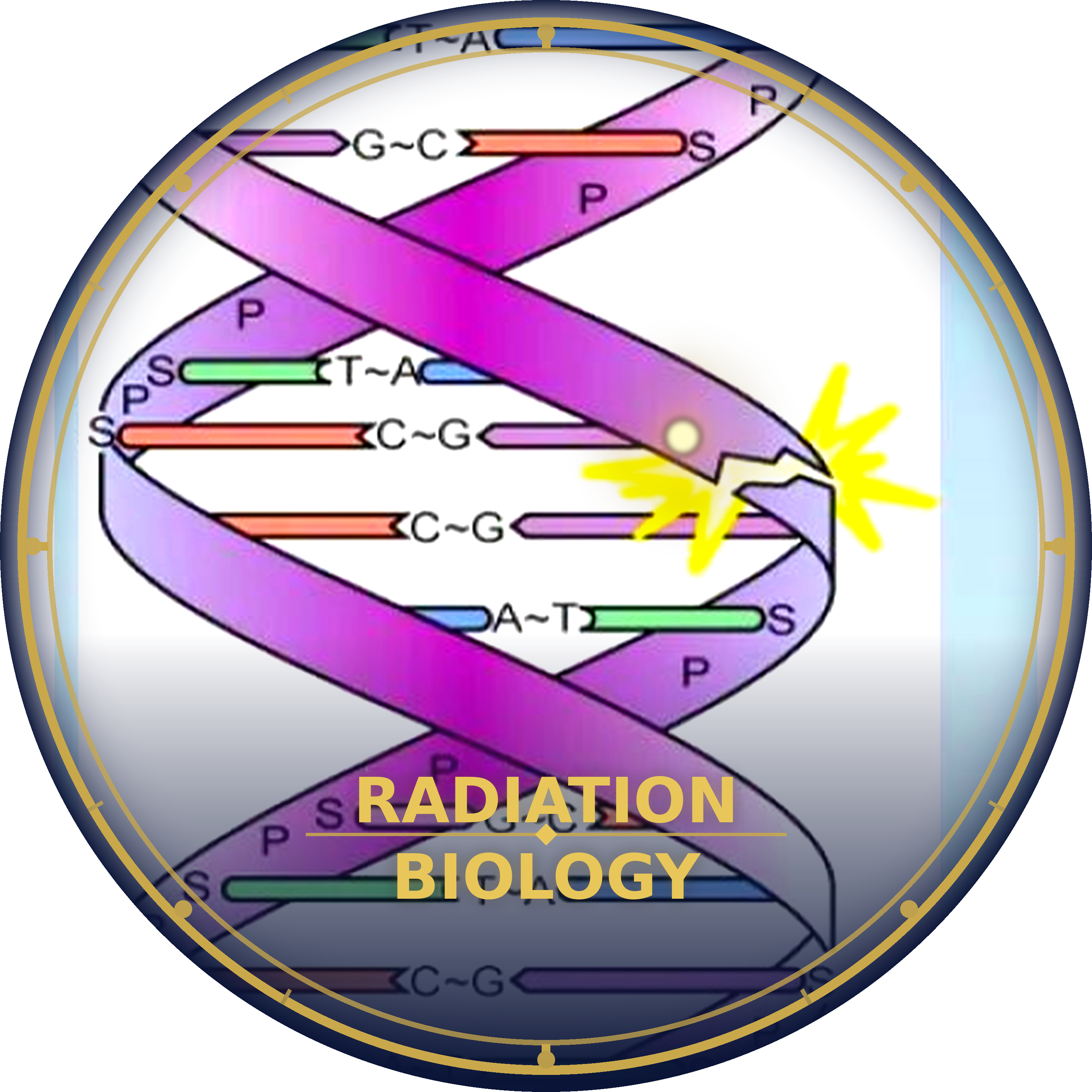
    [Radiation Biology] Ep 2 – X-ray Interactions with Matter

    This episode covers how x-rays are produced in the tube and how the two primary exposure factors — kVp and mAs — shape the energy and quantity of the x-ray beam. Listeners will explore core terminology including primary radiation, exit radiation, transmission, and attenuation, and learn how four key tissue properties — photon energy, thickness, atomic number, and density — determine how much of the beam is absorbed versus transmitted. The episode establishes the foundational relationship: Primary minus Exit equals Attenuation. The five types of x-ray interactions with matter are examined in detail — coherent scattering, Compton scattering, photoelectric absorption, pair production, and photodisintegration — with clear explanation of which interactions are relevant to diagnostic radiography and why. Special emphasis is placed on comparing Compton scattering and photoelectric absorption: how each behaves across energy ranges and atomic numbers, their opposing effects on image contrast and patient dose, and the clinical concept of differential absorption that makes diagnostic radiographs possible. Content is structured to support radiologic technology programs preparing for imaging coursework and ARRT certification review. This episode aligns with the Safety content category — Radiation Physics and Radiobiology subcategory — of the ARRT Radiography Examination Content Specifications. Audio content is adapted from original instructional material developed by Professor Sanjay Arya, M.S., R.T.(R)(MR) for radiologic technology education. Part of the Radiation Biology series — Quantum Imaging Lab. © 2026 Quantum Imaging Lab. All rights reserved.

Showing 41–45 of 45 episodes